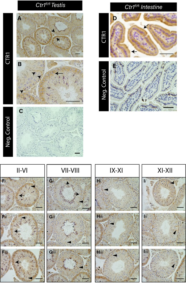
Fig 1. CTR1 is expressed in pachytene spermatocytes and Sertoli cells in a stage specific manner.
Immunohistochemical localization of CTR1 in adult (PND 60) Ctr1fl/fl mouse testis cross sections (A, B). Arrow indicates expression on primary pachytene spermatocytes. Arrowheads indicate SC apical and basal cytoplasm. CTR1 expression on the apical surface of the epithelial cell in the intestine of adult mice indicated by arrows (D). Negative secondary only control of testis (C) and intestine (E) shown. Scale bar = 50 μm. Seminiferous epithelium at stages II-VI displayed the highest CTR1 expression on pachytene spermatocytes (F). Stages VII-XII (G, H and I) display the least CTR1 staining on primary spermatocytes (arrows) but high CTR1 localization on SCs (indicated by arrowheads) within the seminiferous tubule cross sections. Three different tubule cross sections for each specified stage are shown below each grouped of stages (i-iii). Scale bar = 100 μm.

