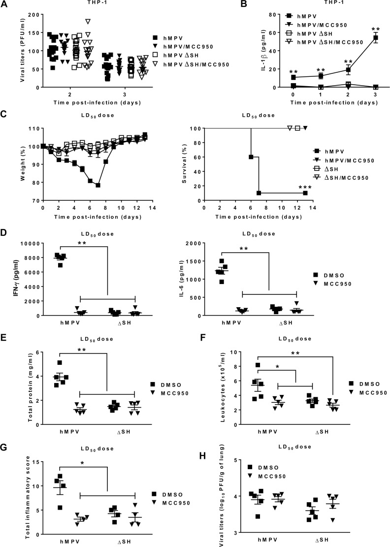
Fig 7. Lack of SH protein promotes attenuated pathogenicity of HMPV.
THP-1 cells were differenciated into macrophages using PMA (100 ng/ml) before treatment with 10 μM of MCC950 or DMSO (control) and then infected or not with HMPV or HMPV ΔSH at a MOI of 0.001. (A) The viral titers were determined by immunostaining. Data were collected from three independent experiments. Values are shown as mean ± S.E.M (unpaired Student t-test, n = 5 per group per experiment). (B) IL-1β levels were measured in the cell supernatants. Values are shown as mean ± S.E.M (Kruskall-Wallis test followed by Dunn’s post hoc, ** P < 0.01 HMPV/MCC950, HMPVΔSH or HMPV ΔSH/MCC950 vs HMPV, n = 5 per group). (C) BALB/c mice were inoculated or not with HMPV at a LD50 dose (5 x 10⁵ PFU per mouse) or HMPV ΔSH in the presence or absence of MCC950 (5 mg/kg). MCC950 treatment was repeated for the next two days (1 time/day). The animals were monitored for survival and weight loss for 14 days after infection and euthanized if humane end points were reached (≤ 20% of initial weight). Values are shown as mean of weight ± S.E.M or average percent survival (Kaplan Meier survial curves, *** P < 0.001 HMPV/MCC950, HMPVΔSH or HMPV ΔSH/MCC950 vs HMPV, n = 10 per group). The lungs and BAL were harvested on day 5 post-infection. (D-F) IFN-γ, IL-6, total protein and leukocyte recruitment were evaluated in BAL (n = 5 per group). (G) histopathology was assessed in the lungs (n = 4 per group). Values are shown as mean ± S.E.M (Kruskall-Wallis test followed by Dunn’s post hoc, * P < 0.05; ** P < 0.01 HMPV/MCC950, HMPVΔSH or HMPV ΔSH/MCC950 vs HMPV). (H) Viral titers were evaluated in lung homogenates. Values are shown as mean ± S.E.M (Kruskall-Wallis test followed by Dunn’s post hoc, n = 5 per group).

