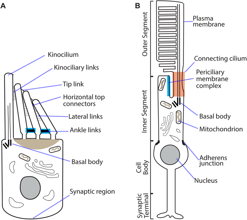
Figure 1:

Structure of an inner ear hair cell and a retinal photoreceptor. (A) A developing cochlear hair cell (e.g., mouse P2–P12) has ankle links (highlighted in blue) located at the base of stereocilia. Lateral links and horizontal top connectors are also present. Tip links connect the tip of short stereocilia to the shaft of longer stereocilia in the adjacent row and are required for mechanotransduction. (B) A rod photoreceptor has an outer segment with discs that contain rhodopsin. Rhodopsin detects light in the form of photons. The photoreceptor inner segment contains mainly organelles required for energy metabolism and protein synthesis. The periciliary membrane complex (highlighted in blue) is located at the apical region of inner segment, surrounding the connecting cilium (highlighted in orange). In order to illustrate the periciliary membrane complex and the connecting cilium, the calyceal processes, which are located outside the two subcellular structures, are not shown. The cell body contains the nucleus, while the synaptic region contains ribbon synapses.
