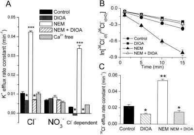
Figure 3.
Signaling cascades involved in the regulation of KCC3 activity. (A) KCC3 activity is inhibited by 20 μM DIOA and stimulated by 0.5 mM NEM, which is sensitive to DIOA (20 μM) inhibition. KCC3 activity is independent of Ca2+ signaling. For Ca2+-free condition, KCC3-transfected cells were preincubated with 50 μM BAPTA-AM for 30 min, and then efflux was measured in Ca2+-free medium containing 1.5 mM EGTA. [BAPTA-AM, 1,2-bis(2-aminophenoxy)ethane-N, N, N′, N′-tetraacetic acid tetrakis(aceto-xymethyl ester)]. (B and C) The Cl− efflux in KCC3-transfected cells is sensitive to DIOA and NEM. Time courses of Cl− efflux (B) from KCC3-transfected cells treated with 20 μM DIOA or 0.5 mM NEM or 20 μM DIOA plus 0.5 mM NEM. Graph shows logarithm of fraction of original intracellular 36Cl− remaining as a function of time. (C) Summary of Cl− efflux rates calculated from the time-course results. To investigate the drug effects on K+ or Cl− efflux, KCC3-transfected cells were preincubated with 0.05% DMSO (control group) or drugs for 15 min at room temperature and then exposed to flux medium in the presence or absence of drugs. Each value in this figure represents mean ± SEM (n = 6). *, P < 0.05; **, P < 0.01; ***, P < 0.005 compared with control group.

