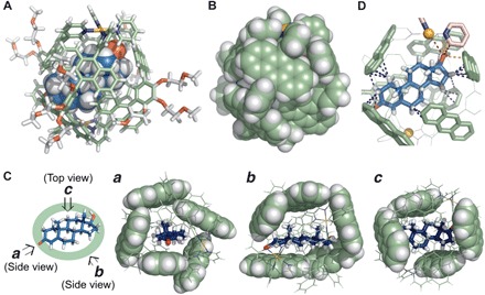
Fig. 3. X-ray crystal structure of 1’•2a.

(A) Space-filling (for 2a) and cylindrical (for 1’) representation and (B) space-filling representation (the peripheral substituents of 1’ are replaced by hydrogen atoms for clarity). (C) Highlighted positions of 2a inside the polyaromatic shell of 1’ (three different views). (D) Highlighted host-guest interactions of 1’•2a in the cavity (blue, orange, and red dashed lines are CH-π, OH-π, and hydrogen-bonding interactions, respectively).
