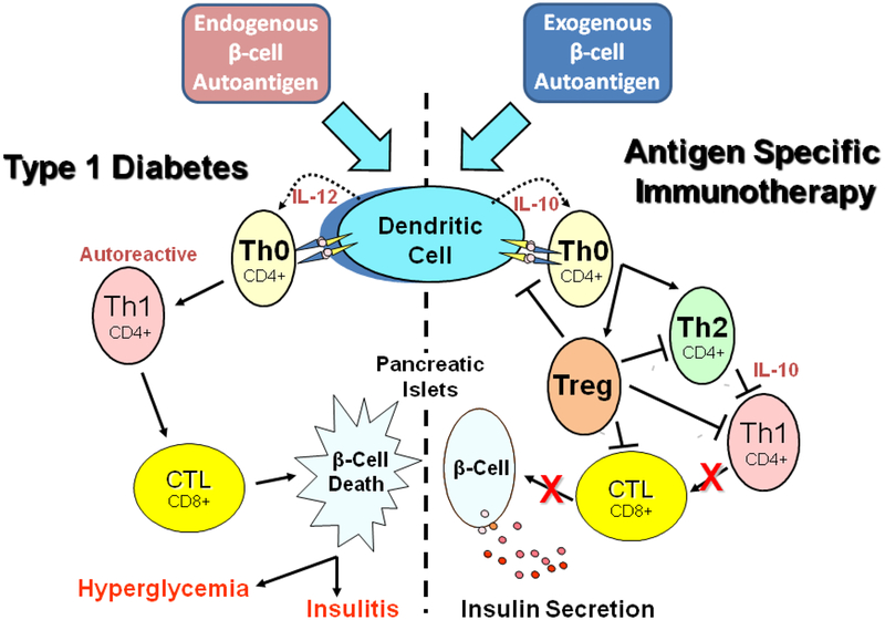
Figure 1.
Basic mechanism of type 1 diabetes inflammation and vaccine action. Antigen presenting cells, specifically dendritic cells, process endogenous pancreatic beta cell autoantigens (Insulin, GAD, etc.) in lysosomal vesicles and transfer peptide fragments of the autoantigen to MHC class II molecules that migrate to the plasma membrane and present the autoantigen to cognate T cell receptors on naïve T helper cells (Th0), left portion of the Figure. At the same time, dendritic cell processing of the autoantigen stimulates biosynthesis and secretion of the inflammatory cytokine interleukin 12 (IL-12) which stimulates the Th0 cells to undergo morphogenesis into autoreactive effector T helper cells (Th1). The autoreactive Th1 cells secrete inflammatory cytokines such as IFN-gamma and IL-2 that stimulate cytotoxic lymphocytes (CTL) to secrete nitric acid, peroxide, and several other inflammatory cytokines that stimulate pancreatic islet inflammation (insulitis). Chronic insulitis results in continual pancreatic beta cell death resulting in increasing insulin deficiency and a progressive increase in blood sugar (hyperglycemia). Oral delivery of small amounts of islet autoantigens (right portion of the Figure) exerts a protective antigen specific therapeutic effect by stimulating dendritic cells to secrete the anti-inflammatory cytokine IL-10 which stimulates naïve cognate Th0 lymphocytes to undergo morphogenesis into anti-inflammatory CD4+ Th2 helper cells that in turn secrete IL-10 which suppresses further development of autoreactive Th1 cells and decreases potential insulitis onset. Alternatively, naive Th0 cells may develop into one of several subclasses of regulatory T cells (Treg), which can block dendritic cell activation, Th2, Th1, and CTL development leading to prevention of insulitis, continued insulin secretion, and maintenance of immunological homeostasis.

