Fig. 7.

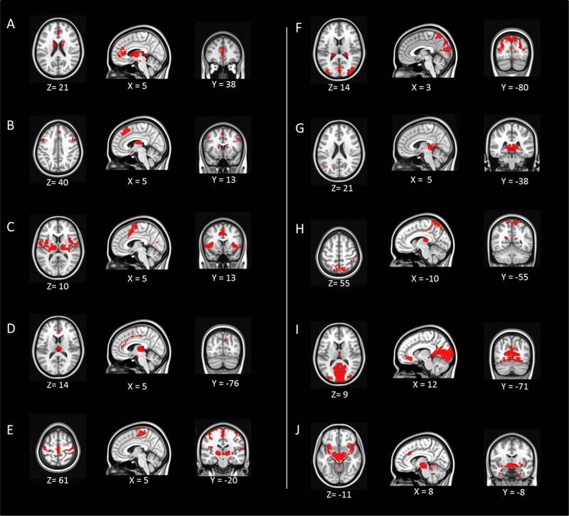
The resting-state functional connectivity of each thalamic cluster. Each panel starting from A to J corresponds to Cluster 1–10. The map is in MNI space. The x, y, z coordinates of each image is shown (left = left).

The resting-state functional connectivity of each thalamic cluster. Each panel starting from A to J corresponds to Cluster 1–10. The map is in MNI space. The x, y, z coordinates of each image is shown (left = left).