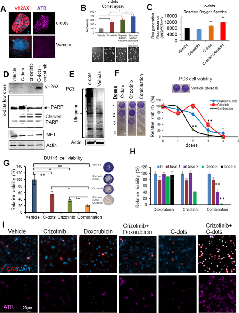
Figure 7. Carbon nanodots sensitize PCa cells to MET inhibitor.

(A) C-dots induce DNA damage response in DU145 cells at dose of 0.1mg/ml. (B-C) C-dots induce more DNA damage response and ROS generation in PC3 cells with MET inhibitor crizotinib. Cells were treated by c-dots at 0.1mg/ml with or without 10μM crizotinib followed by comet or ROS assay. (D) Western blot showing C-dots at low dose of 0.025 mg/ml enhance DNA damage response, cleaved PARP, and decreased MET levels in PC3 cells with MET inhibitor crizotinib. (E) C-dots induce high molecular weight protein ubiquitination in PC3 cells. (F-H) C-dot or DOX increases efficacy of MET inhibitor on PCa cell growth. (I) C-dot or DOX increases efficacy of MET inhibitor on DNA damage response. Data are representative of averages+ SD (standard deviation).
