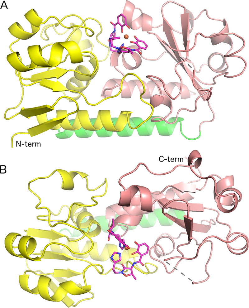
Figure 2.
Ribbon representation of BauB bound to the Acb2•Fe complex. (A) Highlight of the two domains and the α helix that joins them (N-domain, yellow; C-domain pink; α helix green) (B) Orthogonal view, rotated approximately 90° around the horizontal axis. The single gap in the protein between residues 235 and 239 is indicated with the dashed line. The Acb molecules are shown in stick representation with magenta carbon, red oxygen, and blue nitrogen atoms.

