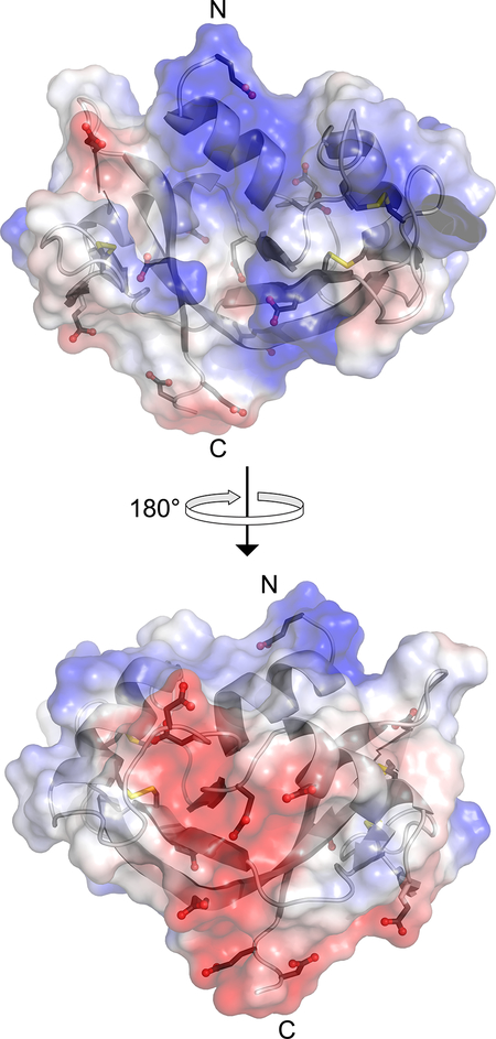
Figure 1.
Surface electrostatic potential of human RNase 1 (blue, positive; red, negative). The side chains of the 6 aspartate, 6 glutamate, and 4 cystine residues are shown explicitly. The image was created with the program PyMOL from Schrödinger (New York, NY) and Protein Data Bank entry 1z7x, chain X.22

