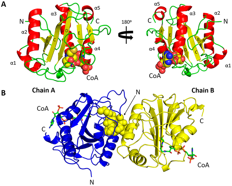
Figure 1.
Crystal structures of PA3944 in complex with CoA. (A) Monomer assigned to an orthorhombic space group (PDB entry 6edv). α-Helices (red) are labeled from α1 to α5, β-strands (yellow) labeled from A to F, and loops colored green. CoA is shown as spheres. (B) The N-terminus (spheres) of one monomer (PDB entry 6edd, chain B, yellow) is bound to the active site of the other monomer (PDB entry 6edd, chain B, blue). CoA is shown as green sticks. The N- and C-termini are labeled as N and C, respectively, in both panels.

