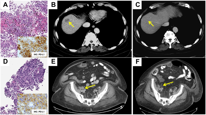
Figure 1: Histopathology & Imaging.

Representative histology of a liver biopsy showing metastatic cutaneous melanoma involving the liver (A, Case 1) and CT imaging showing response to therapy with Pembrolizumab (B: 2.5cm × 1.6cm on Day 1; C: 0.7cm × 0.6cm on Day 313). Representative immunohistochemistry for PD-L1 is depicted as well (A, inset). Histology of an inguinal mass biopsy showing involvement by metastatic mucosal melanoma (D, Case 2) and corresponding immunohistochemistry for PD-L1 (D, inset). CT imaging showing resolution of the lesion following therapy with Pembrolizumab (E: 2.4cm × 1.9cm, Day 256; F: no appreciable adenopathy, Day 1050) is depicted. Arrows demonstrate the lesion of interest.
