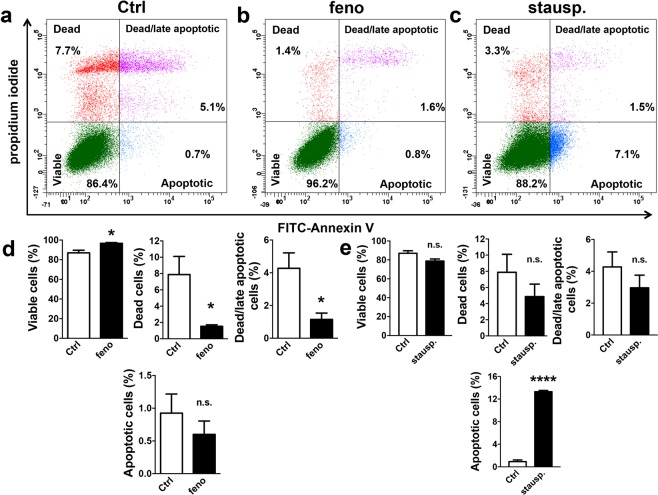
Figure 2.
Fenofibrate did not induce apoptosis in MS1 VEGF angiosarcoma cells. Data were generated in MS1 VEGF angiosarcoma cells by flow cytometry. (a–c) Example experiment showing the proportion (%) of dead, early apoptotic, or dead/late apoptotic cells after treatment with 50 μM fenofibrate (feno) or 1 μM staurosporine (stausp). Staining for FITC-conjugated Annexin V was used as a marker for early apoptosis whereas propidium iodide (PI) staining was used as a marker for cell death. (d,e) Mean data for experiments exemplified in a-c (n = 4, fenofibrate data set; n = 3, staurosporine data set). Data were analysed using a Student’s t-test. *P < 0.05; ****P < 0.0001; n.s, not significant.

