
Fig. 2.
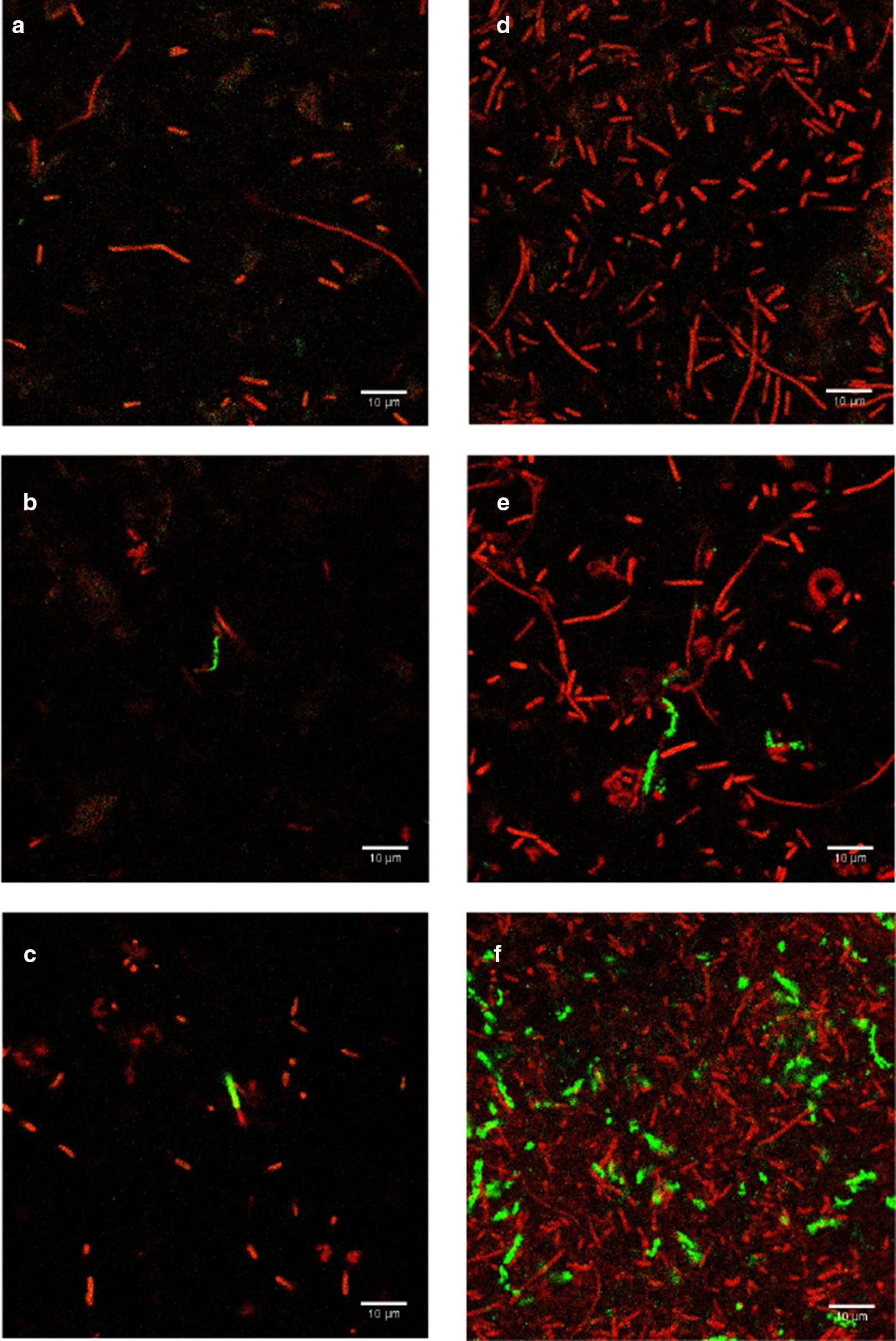
Representative fluorescent images of E. coli BL21 102 CFU/ml after 4 h enrichment in TSB: a no phage infection, b after 15 min of T7-ALP infection, c after 30 min of T7-ALP infection. Fluorescent images of E. coli BL21 103 CFU/ml after 4 h enrichment in TSB: d no phage infection, e after 15 min of T7-ALP infection, f after 30 min of T7-ALP infection