
Figure 2.
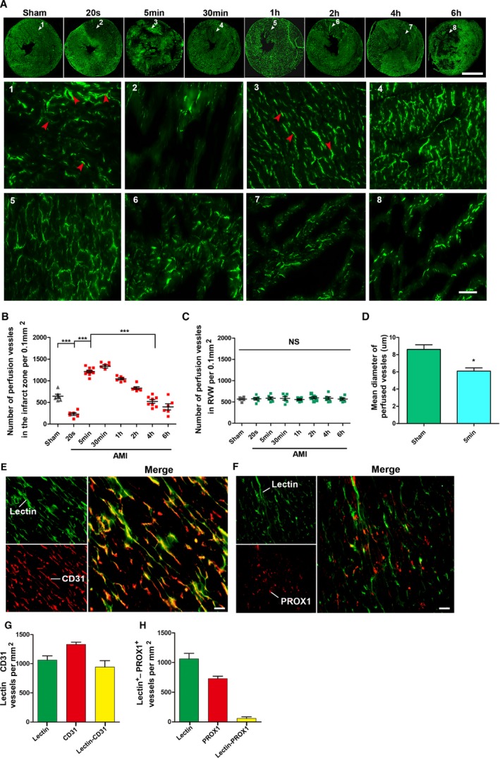
Distribution of CCMR vessels in rat hearts (the left ventricular myocardium). A, Lectin‐FITC perfusion experiment was conducted in rats to mark vessels with blood perfusion at 20 seconds, 5 minutes, 30 minutes, 1 hour, 2 hours, 4 hours, and 6 hours ligation of the left anterior descending coronary artery; bar (upper)=5 mm, bar (lower)=50 μm. White arrows are the selected representative area; red arrows indicate perfused microvessels labeled by lectin‐FITC (n=6–9). B and C, Quantification of vessels with blood perfusion in infarction zone (left ventricular wall) and relative normal region (right ventricular wall; RVW); values are means±SD; *P<0.05 vs the sham group; ***P<0.001 vs the indicated groups NS; P>0.05 vs the sham group. D, Quantification of mean diameter of perfused vessels in the sham and 5‐minute group. E, Light microscopy analyses coexpression of CD31 (red) in lectin‐labeled vessels (green) in 5‐minute group cardiac sections (bar=50 μm; n=7). F, Light microscopy analyses coexpression of PROX1 (red) in lectin‐labeled vessels (green) in 5‐minute group cardiac sections (bar=50 μm). G and H, Quantification of CD31+ Lectin+ vessels and PROX1+Lectin+ vessels. CCMR indicates coronary collateral microcirculation reserve; NS, not significant; PROX1, prospero homeobox 1.