
Figure 2.
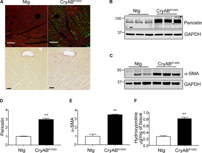
Protein markers of fibrosis in mutant αB‐crystallin (CryABR 120G) mice. A, Periostin (green) and Sirius Red–stained left ventricle sections from 4‐month‐old nontransgenic (Ntg) and CryABR 120G hearts. In periostin immunohistochemistry sections, cardiomyocytes are labeled with troponin I (red) and nuclei with DAPI (blue). The scale bar is 50 μm for the upper panels and 100 μm for the lower panels. B, Expression of the fibrosis markers, periostin and (C) α‐smooth muscle actin (α‐SMA), in left ventricles derived from 4‐month‐old Ntg and CryABR 120G hearts. D, Quantitative analysis of the periostin and (E) α‐SMA western blots: shown are the ratio values using GAPDH to normalize loading variation (n=6). F, Hydroxyproline levels derived from 4‐month‐old left ventricles (n=5). **P<0.001 vs Ntg control. DAPI indicates 4′,6‐diamidino‐2‐phenylindole; GAPDH, glyceraldehyde 3‐phosphate.