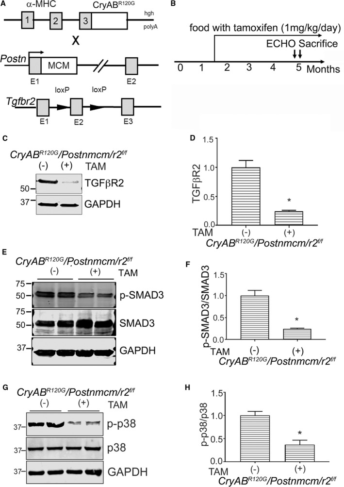
Figure 8.

Generation of myofibroblast specific transforming growth factor β receptor II gene (Tgfbr2) (r2)–ablated mice. A, Schematic representation of the different mouse lines used. Mice with cardiomyocyte‐specific mutant α‐B‐crystallin expression (CryABR 120G) were crossed with Tgfbr2‐loxP–containing gene targeted lines and those double transgenic (Tg) lines further crossed with mice containing the periostin locus (Postn) with a tamoxifen (TAM) regulated Mer‐Cre‐Mer cassette (Postnmcm) inserted into exon (E1). B, Experimental scheme where mice were fed a TAM diet starting from 6 to 8 weeks until they were euthanitized. C and D, Western blot and quantification for Tgfβr2 protein in purified fibroblasts (n=6 separate, total hearts). E and F, Expression and quantification of SMAD3 (phospho [p]‐SMAD3/SMAD3) and (G and H) p38 signaling (p‐p38/p38) in purified fibroblasts isolated from hearts of 3‐month‐old experimental cohorts fed with TAM chow (TAM+) or normal chow (−) (n=4–6 separate, total hearts). *P<0.05 vs uninduced Cre Cry ABR 120G /Postnmcm/r2 f/f animals. GAPDH indicates glyceraldehyde 3‐phosphate; α‐MHC, α‐myosin heavy chain; SMAD3, small mothers against decapentaplegic homolog 3.
