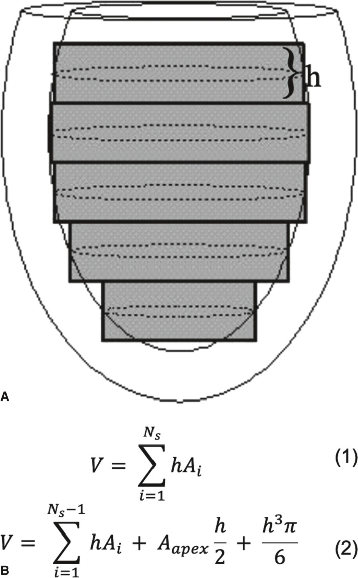
Figure 1.

Composite midpoint integration methods for parallel short‐axis image data. A, A diagram of an elliptical virtual left ventricle in a slightly oblique side view with short‐axis cut‐planes of thickness h indicated by gray rectangles. The in‐plane endocardial contours are depicted as dotted‐line ellipses. B (top), Gives the equation for the basic composite midpoint integration method as sum over all (N s) cross‐sectional slice areas (A i). B (bottom), Gives a variant from Wyatt et al17 intended to correct for curvature effects at the apex. The sum is over all slices except the apex, then adds half the apex volume plus the volume of a hemisphere of radius equal to half the slice thickness.
