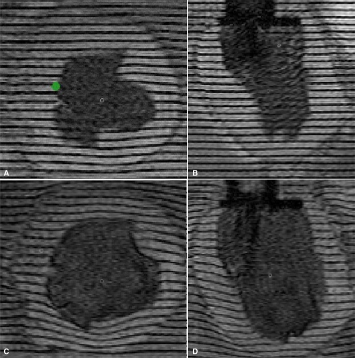
Figure 5.

Representative high‐resolution magnetic resonance images of an isolated canine left ventricle (LV) undergoing passive inflation. A and B, Representative short‐axis and long‐axis images in the experimental end‐systolic state. C and D, The same image planes but in the experimental, physiologic end‐diastolic state. The signal decrease in the LV cavity results from deuterium (D2O) added to the water filling the balloon. The Teflon mitral valve collar and nozzle assembly is apparent as a darker‐appearing slab in the top center of the long‐axis images. The green dot in (A) represents the middle of the septal wall.
