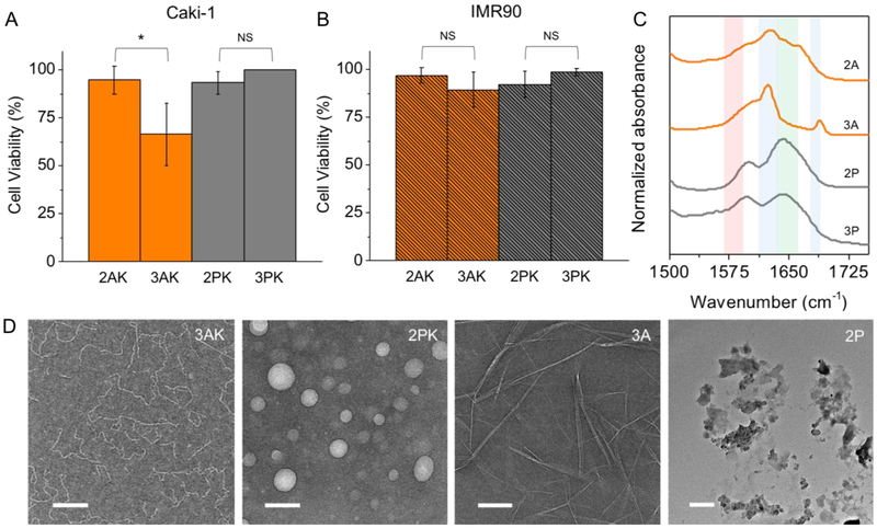
Figure 4.
Biocompatibility of peptide nanostructures. Cell viability of human renal cancer cell line, Caki-1, with high expression of MMP-9 (A), and non-cancerous human fibroblast, IMR90, with normal expression of the enzyme (B), incubated with 1 mM peptides for 72 hrs. The peptides are non-toxic to both cell lines, except for 3 AK which decreased the cell viability of Caki-1 to 66%. (C) FTIR spectra of the post-cleavage peptides show that only 3 A forms anti-parallel β-sheet peptide fibers which causes the cytotoxic effects in Caki-1 only. (D) TEM images of 3 AK and 2 PK show formation of worm-like and spherical micelles, respectively, and the post-enzymatic products, 3 A and 2 P which form rigid fibers or random aggregates, respectively. Scale bar 100nm.

