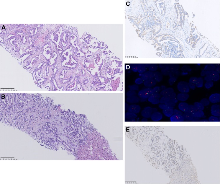
Figure 4.
Analysis of the primary left breast and liver lesion tissue.
Notes: (A) H&E; (C) IHC, HER2 2+; (D) FISH, HER2-; analysis of the primary breast lesion. (B) H&E; (E) IHC, HER2-; analysis of metastatic liver lesion. H&E stained images are depicted at 100× magnification.
Abbreviations: FISH, fluorescence in situ hybridization; HER2, human epidermal growth factor receptor 2; IHC, immunohistochemistry.

