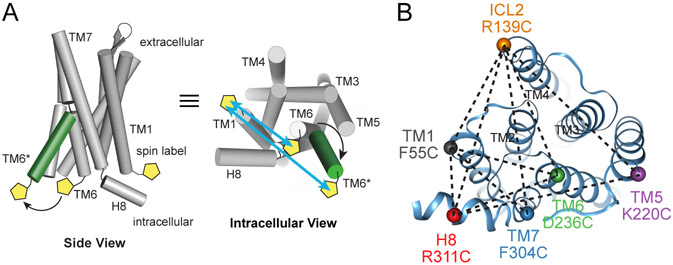
Figure 1: DEER Distance Measurements of the AT1R.
(A) Site-directed spin labeling of AT1R intracellular regions with the nitroxide-containing V1 side chain allows the detection of ligand-dependent conformational changes by DEER spectroscopy, as shown schematically for TMs 1 and 6.
(B) AT1R labeling strategy indicating six labeling sites and their ten pairwise combinations (dotted lines), shown on the crystal structure of AT1R bound to the candesartan precursor ZD7155 (PDB: 4YAY).

