Fig. 2.
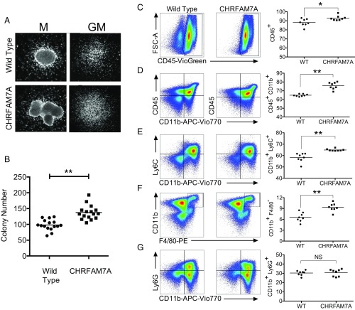
MethoCult identifies an increase in colony-forming activity and bias to monocyte lineage. (A) Morphology of colonies from wild-type and CHRFAM7A-transgenic mice. The appearance of macrophage (M) and granulocyte-monocyte (GM) colonies expanded from 3 × 10³ bone marrow cells from wild-type and CHRFAM7A-transgenic mice in GM differentiating MethoCult is morphologically indistinguishable. Cells were examined under phase-contrast microscopy and photographed. (B) Colony numbers in GM differentiating MethoCult. After 7 d in MethoCult culture, the numbers of colonies that arose after plating 3 × 10³ wild-type or CHRFAM7A-transgenic bone marrow cells per dish were determined. Differences in 16 replicates performed from 8 donor mice were analyzed by unpaired, two-tailed t test (P < 0.01). The horizontal line shows the mean. (C–G) Flow cytometry of bone marrow cells differentiated in vitro. After the exclusion of debris, doublets, and nonviable cells (SI Appendix, Fig. S10), flow cytometry was used to identify and measure changes in myeloid differentiation including CD45+ (C) and CD45+CD11b+ (D) leukocytes, and CD11b+Ly6C+ monocytes (E) and F4/80+CD11b+ macrophages (F). There was no difference in CD11b+Ly6G+ granulocytes (G). Differences in eight mice were analyzed by unpaired two-tailed t test (*P < 0.05, **P < 0.01; NS, not significantly different). The horizontal line shows the mean.

