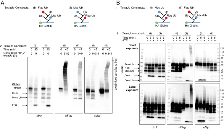
Fig. 3.
Fates of proximal and distal Ub moieties in a tetra-Ub chain. (A, 1) Sketches of differentially tagged tetra-Ub HA-α-globin: (i) tetra-Ub HA-α-globin with distal Flag-Ub and proximal Myc-Ub and (ii) tetra-Ub HA-α-globin with distal Myc-Ub and proximal Flag-Ub (also see Fig. 2A, constructs 15 and 16, respectively). (A, 2) Differentially tagged tetra-Ub HA-α-globin constructs were incubated for the indicated times in the presence of crude fraction II and ATP. Following SDS/PAGE, reactions were analyzed by Western blot analysis with αHA (Left), αFlag (Middle), and αMyc (Right). Quantification of the Flag-Ub- and Myc-Ub conjugates is presented as the ratio of the Flag or Myc signal in the generated conjugates at 45 min (minus the signal at time 0) relative to the Flag or Myc signal in the tetra-Ub HA-α-globin at time 0. (B, 1) Sketches of differentially tagged tetra-Ub HA-α-globins: (i) tetra-Ub HA-α-globin with distal Myc-Ub and proximal Flag-Ub and (ii) tetra-Ub HA-α-globin with distal Flag-Ub and proximal Myc-Ub (also see Fig. 2A, constructs 16 and 15, respectively). (B, 2) Differentially tagged tetra-Ub HA-α-globin constructs were incubated for the indicated times in the presence of fraction II with and without ATP. Following SDS/PAGE, reactions were analyzed by Western blot analysis as described in A, 2. The same image is shown with short (Upper) and long (Lower) exposures.

