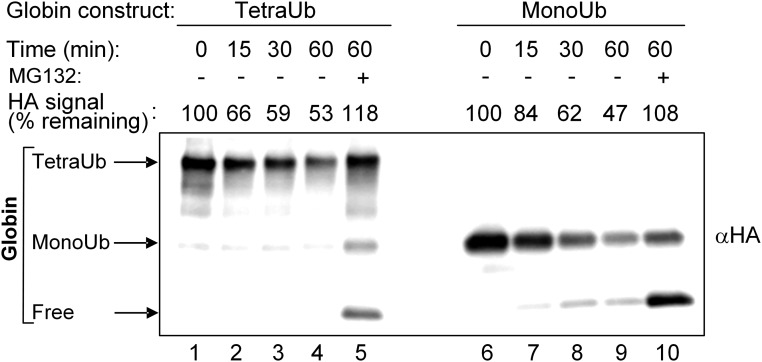
Fig. 6.
Similar degradation of tetra-Ub HA-α-globin and mono-Ub HA-α-globin by purified 26S proteasome. Tetra-Ub HA-α-globin with distal Flag-Ub and proximal Myc-Ub and mono-Ub HA-α-globin with Flag-Ub (Fig. 2A, constructs 15 and 11, respectively) were incubated for the indicated times in the presence of purified human 26S proteasome (1.5 μg). MG132 (100 μM) was added as indicated. Following SDS/PAGE, reactions were analyzed by Western blot analysis using αHA. Quantification of degradation at the different time points was calculated as described in Fig. 5.

