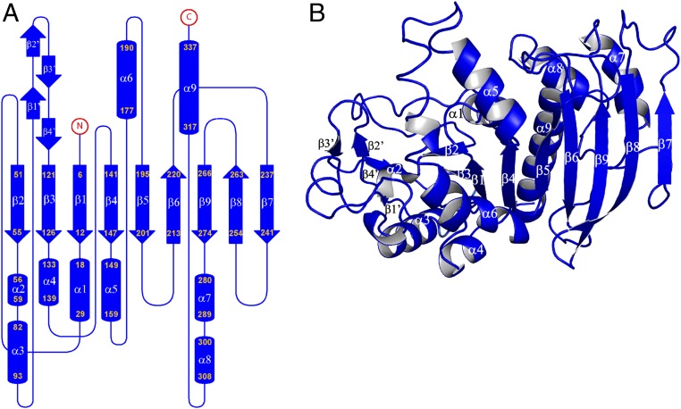
Fig. 2.
Crystal structure of the CrPRK monomer. (A) Topology diagram: The monomer is composed of a mixed β-sheet of nine strands, nine α-helices, and four additional small β-strands indicated by β′. (B) Representation of the structure of the monomer: The central β-sheet is sandwiched between helices α3, α4, and α6 and helices α1, α7, α8, and α9. Strand β7 is located in the dimer interface, whereas the four additional β-strands (β1′ to β4′) form the edge of the dimer.

