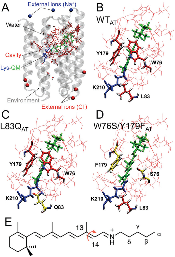
Figure 1.
QM/MM models. (A) General structure of the ASR model. The protein environment is colored in grey with external counterions in blue (Na+) and red (Cl−). The region hosting the retinal chromophore is colored in red with the Lys210-chromophore system in blue-green. Chromophore hosting region for the all-trans isomers of WT (ASRAT), L83Q and W76S/Y179F are given in part (B), (C) and (D) respectively. The variable cavity residues 76, 83 and 179 are shown in tube representation. (E) The all-trans retinal chromophore. The curly arrow indicates the C13=C14 isomerizing double bond and the greek letters the atoms of the Lys210 side chain.

