
Figure 3. Resistive-pulse device and pulse-amplitude measurements.
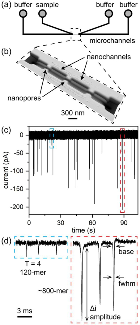
(a) Two V-shaped microchannels are connected through a series of nanochannels and nanopores, indicated with the dashed box. (b) Atomic force microscope (AFM) image of the nanochannels and nanopores. The nanopores are 100 nm wide, 100 nm deep, and 280 nm long. (c) Typical current trace of an assembly reaction of 5 μM Cp dimer with 20 μM HAP-TAMRA and 300 mM NaCl. The current baseline (~17 nA) was subtracted from the signal. Each of what appears as a single pulse on the timescale of the plot in panel (c) consists of (d) three discrete pulses. In panels (c) and (d), the blue dashed line indicates detection of an individual T = 4 capsid, and the red dashed line indicates detection of one of the largest protein particles formed. The arrows in panel (d) indicate the pulse width at a fixed position (3σ or 4σ) below the baseline (base) and pulse width at half-height (i.e., full width at half-maximum, fwhm).