
Figure 6. Morphological information extracted from nanochannel data.
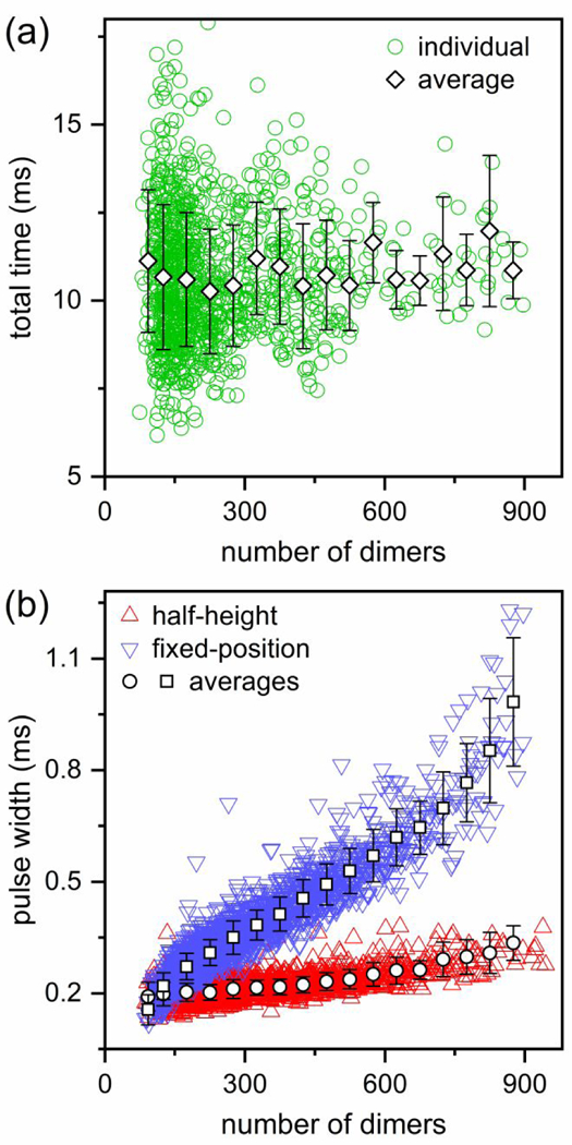
(a) Variation of the total transit time of each particle through the three pores and two pore-to-pore nanochannels with number of dimers. The total time does not change with particle size which indicates that the particles of different sizes have similar electrophoretic mobilities. (b) Variation of average pulse width with number of dimers. The pulse width measured at half-height increases only slightly, whereas the width measured at a fixed position (3σ or 4σ below the baseline current) increases significantly with increased number of dimers. Particles were assembled from 5 μM Cp dimer with 20 μM HAP-TAMRA and 300 mM NaCl in 50 mM HEPES (pH 7.5).