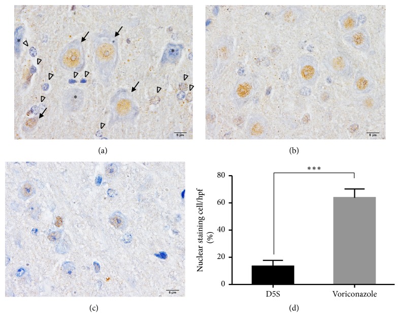
Figure 4.
Immunohistochemical staining of Nrf-2 in the neurons. Nrf-2 labeling was expressed in the nucleus of the neuronal ((a); arrow) and supporting ((a); arrowhead) cells. However, the nucleolar expression was decreased in some of the abnormal neurons ((a); ∗). The comparison of Nrf-2 level between the brain without (b) or with (c) treatment was shown in (d). ∗∗∗: p value < 0.0001.

