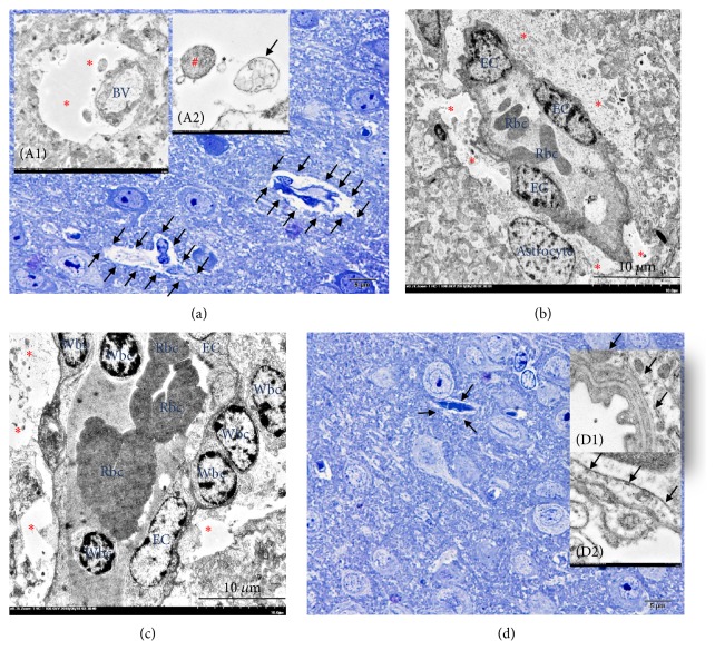
Figure 6.
Ultrastructure of cytogenic oedema in scedosporiosis-induced cerebral abscess. In the brain with abscess, the presence of cytogenic oedema was frequent. It was characterized by astrocyte foot process dilatation where the perivascular area was distended ((a), (b) and (c); arrow and (A1); ∗) and contained with intracellular materials such as mitochondria ((A2); #) or multivesicular bodies ((A2); arrow). In the brain without abscess, the presence of the perivascular area was less than in abscessed brain. The perivascular area ((d); arrow) and the endothelial lining cell were intact ((D1); arrow) when compared to the brain with abscess represented by vacuolated endothelial cell ((D2); arrow).

