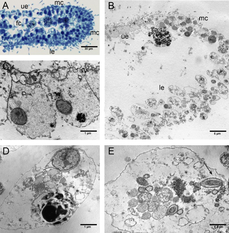
Figure 6. Fine morphology of EtOH Placozoa.
(A) Body cross-section showing the upper and lower epithelium, the margin and the inner fiber cells. Note the considerable width of the margin (optical microscopy). (B) Body cross-section showing the upper and the lower epithelium and the thick margin composed of a group of small cells. (C) Detail of two columnar cells of the upper epithelium. (D) Single fiber cell of the inner three-dimensional syncytium showing typical organelles. (E) Fiber cells: note one bacterium (arrow) inside the cisternae of the rough endoplasmic reticulum. (A): optical microscopy. (B)–(E): TEM microscopy. fc, fiber cells; le, lower epithelium; mc, marginal cells; ue, upper epithelium.

