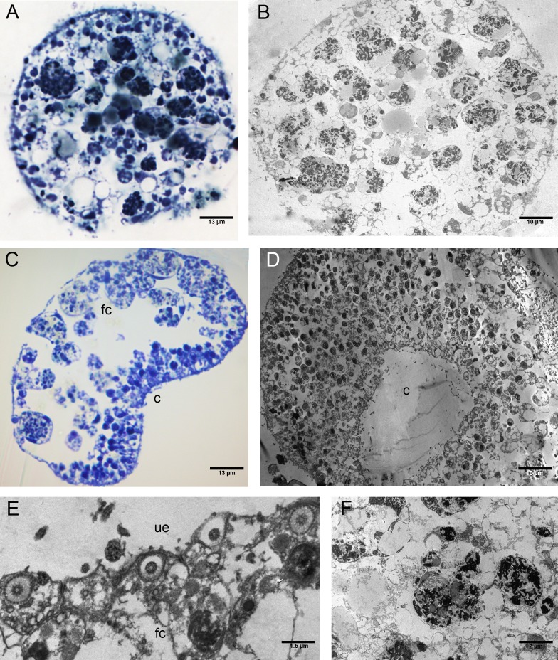
Figure 7. Fine morphology of P+EtOH Placozoa.
Frontal semithin (A) and thin (B) section showing the rounded shape and regular margins of the body. Semithin (C) and thin (D) sections showing the cavity formed by lifting of the lower epithelium in the central part of the body. The majority of the fiber cells are located close to the upper epithelium. (E) Detail of the upper epithelium in which the small cuboidal cells are clearly visible. (F) Thin sections of fiber cells: weakly electron-dense lipid granules and strongly electron-dense proteinaceous material are visible. (A), (C): optical microscopy. (B), (D), (E), (F): TEM microscopy. fc, fiber cells; c, cavity; ue, upper epithelium.

