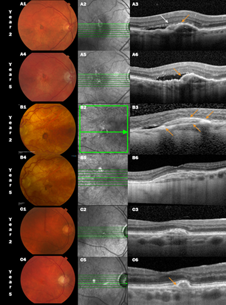
Figure 2:
Color fundus photograph (CFP, left), infrared scanning laser ophthalmoscopic (IR, center) and spectral domain optical coherence tomographic (SDOCT, right) images of non-fibrotic scar (NFS) in three CATT subjects at year 2 and 5. A small pigment encircled NFS is seen at 2 years (A1, B1 and C1) diminishes at year 5 (A4, B4 and C4). Pigmented areas appear bright on the IR images (A2–C2). SDOCT A3 and A6 show compact subretinal hyperreflective material (SHRM, white arrow) over the fibrovascular pigment epithelial detachment (PED) with adjacent subretinal fluid (SRF), however, there is a hyperreflective layer extending from the retinal pigment epithelium (RPE) across part of the inner border of the SHRM (orange arrow). By year 5, the SHRM is now encased in the RPE layer contiguous with the inner border of the PED, subretinal fluid persists with focal sites of greater penetration of OCT signal into the choroid. B1, B2 and B3 show a NFS at 2 years in another study eye along with a similar appearance as in the previous eye (A3) but with an even more prominent region of heaped up hyperreflectivity corresponding to the sites of the dark pigment ring and a second “RPE layer” extending across the SHRM. However the SHRM has either resolved or cannot be distinguished from the layered reflectance in the fibrovascular PED (B6) and the SRF has resolved. In the third example C1–C3, the year 2 compact SHRM (C3) contracts further at year 5 with a more pronounced hyperreflective RPE layer over the lesion (C6).

