FIG 7.
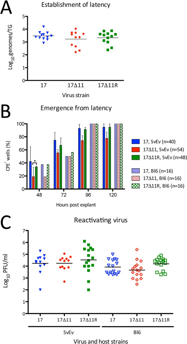
Establishment of and reactivation from latency. Trigeminal ganglia were collected from wild-type 129SvEv and C57BL/6J mice 28 dpi with 2 × 10⁴ PFU/eye of strain 17, strain 17Δ11, or strain 17Δ11R. (A) DNA extracted from latently infected 129SvEv TGs was analyzed for viral thymidine kinase copy number by qRT-PCR. Data were normalized by comparison to data from a single-copy mouse adipsin gene. Data are expressed as numbers of genome copies per individual TG. (B) 129SvEv and C57BL/6J TG explants were plated with indicator Vero cells for 24 h during the interval 48 to 72 hpe. Wells were scored as CPE+ or CPE− at 48, 72, and 96, and 120 h. Data shown represent levels of CPE+ wells as a percentage of total wells (shown in legend) within each infection cohort. *, P < 0.05 (two-way ANOVA and Bonferroni posttests). (C) At 72 hpe, TG from 129SvEv (SvEv) and C57BL/6J (Bl6) mice were homogenized and titers in the homogenate were determined on Vero cells. In panels A and C, bars indicate the mean titers, which were not significantly different (P > 0.05 [one-way ANOVA]).

