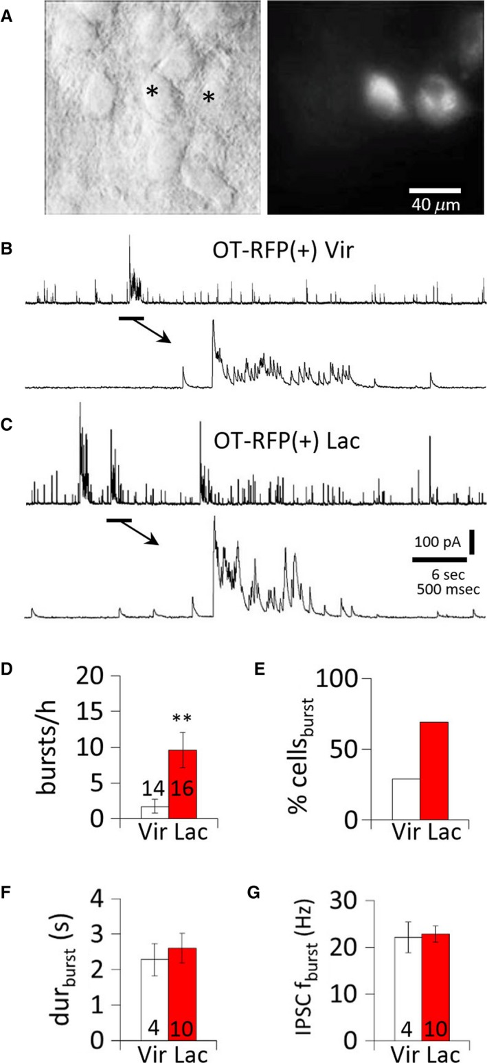
Figure 2.

Upregulation of IPSC bursts in OT MNCs during lactation. (A) OT MNCs viewed in a brain slice under IR‐DIC (left) and epifluorescence (right). *, RFP(+) neurons. (B) Representative IPSC burst recorded in an OT RFP(+) MNC from a non–lactating rat. (C) Representative IPSC bursts recorded in an OT RFP(+) MNC from a lactating rat. (D) Average incidence of bursts of IPSCs (bursts/h) in OT‐RFP(+) MNCs from virgin and lactating rats. (E) Percent of OT‐RFP(+) MNCs displaying IPSC bursts (% cellsburst) in virgin and lactating rats. (F) Mean IPSC burst duration in OT‐RFP (+) MNCs from virgin and lactating rats. (G) Mean IPSC frequency in IPSC bursts in OT‐RFP(+) MNCs from non–lactating and lactating rats. **, P < 0.01.
