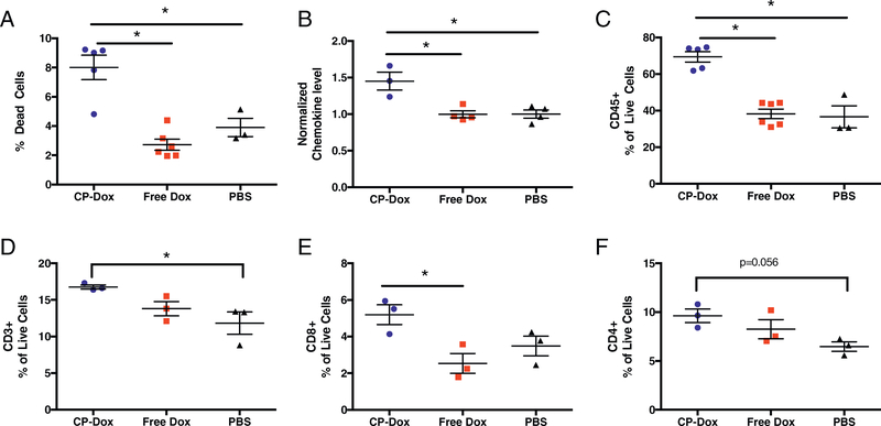
Fig. 3.
CP-Dox treatment increases tumor cell death and intratumoral leukocyte infiltration. One week after treatment of 4T1 mammary carcinoma with CP-Dox, Free Dox, or PBS, tumors were processed to a single-cell suspension and examined by flow cytometry or homogenized and analyzed for chemokine levels. (A) Percentage of dead cells (n = 5 CP-Dox, 6 Free Dox, 3 PBS). (B) Normalized and averaged values for 7 chemokines (raw data found in Supplementary Fig. 1) (n = 3 CP-Dox, 4 Free Dox, 4 PBS). (C) Leukocyte (CD45+) as a percentage of Live cells (n = 5 CP-Dox, 6 Free Dox, 3 PBS). (D-F) CD45+ cells were then gated for quantification of (D) T cells (CD3+), then (E) CD8+ and (F) CD4+ cells as a percentage of live cells (n = 3 CP-Dox, 3 Free Dox, 3 PBS for D-F). Data analyzed by ANOVA and Tukey’s post-hoc. *p < 0.05.

