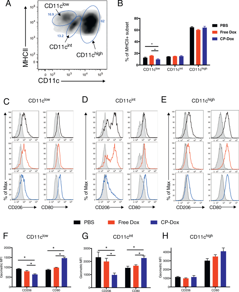
Fig. 6.
Treatment with CP-Dox alters the phenotype of mononuclear phagocytes in 4T1 mammary carcinoma. Mice were inoculated with 4T1 mammary carcinoma and treated with drug as described earlier. One week after drug treatment, cells were processed to a single cell suspension and analyzed by flow cytometry. (A) Flow cytometry plot showing CD45+/ CD11b + / Ly6G− / IA/IE+ myeloid cells (TAMs) for a PBS-treated mouse, displayed as CD11c vs. IA/IE (MHCII), revealing three subsets of cells based on their CD11c expression. (B) Breakdown of each subset as a percentage of IA/IE+ cells for each treatment group (n = 5 CP-Dox, 5 Free Dox, 3 PBS). (C–E) Flow cytometry histograms for the CD11clow, CD11cint and CD11chigh subsets, respectively for CD206 and CD80 expression for treatment with PBS (black), free Dox (red) or CP-Dox (blue). (F–H) Quantification of CD206 and CD80 expression for the (F) CD11clow, (G) CD11cint, and (H) CD11chigh subset for different treatments (n = 5 CP-Dox, 5 Free Dox, 3 PBS). (*p < 0.05). (For interpretation of the references to colour in this figure legend, the reader is referred to the web version of this article.)

