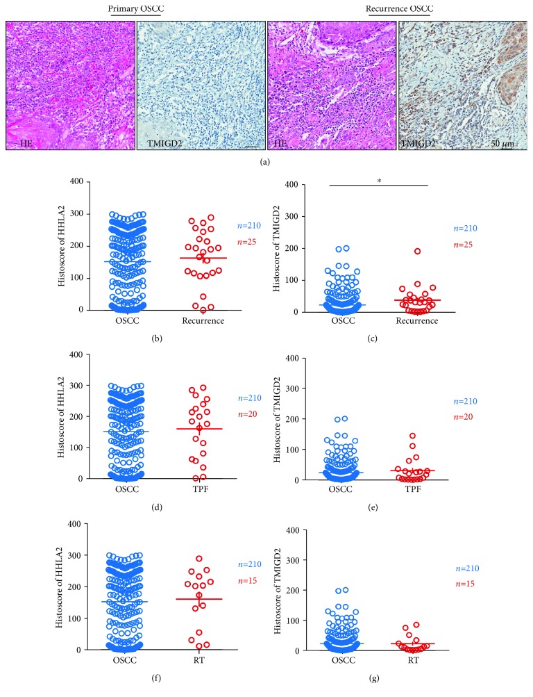
Figure 3.
The TMIGD2 expression level was increased in recurrent OSCC. (a) Hematoxylin and eosin (HE) staining with TMIGD2 immunostaining of primary OSCC and recurrent OSCC. (b) There was no significant difference in the HHLA2 expression levels between primary OSCC and recurrent OSCC. (c) The TMIGD2 expression level was significantly increased in recurrent OSCC compared with primary OSCC. (d) There was no significant difference in the HHLA2 expression levels between primary OSCC and OSCC after TPF therapy. (e) There was no significant difference in the TMIGD2 expression levels between primary OSCC and OSCC after TPF therapy. (f) There was no significant change in the HHLA2 expression level in OSCC after radiotherapy compared with primary OSCC. (g) There was no significant change in the TMIGD2 expression level in OSCC after radiotherapy compared with primary OSCC.

