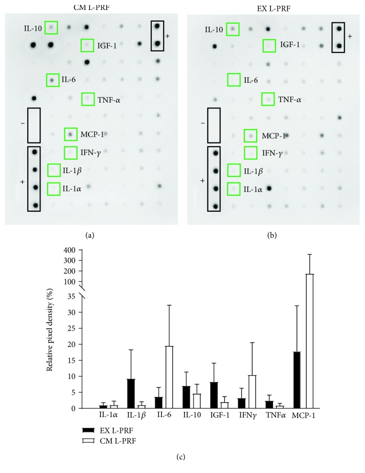
Figure 2.
Protein release profile of EX and CM L-PRF. Representative antibody array of the proteins released from CM L-PRF (a) and EX L-PRF (b) (n = 4). Semiquantitative analysis using relative pixel density (c) was performed to compare relative protein levels between CM and EX L-PRF. Both CM L-PRF (a) and EX L-PRF (b) contained several inflammatory mediators, including IL-1α, IL-1β, IL-6, IL-10, IGF-1, IFN-γ, TNF-α, and MCP-1. None of these mediators were significantly more abundant in either L-PRF fraction. Data are presented as mean ± SEM.

