Figure 4.
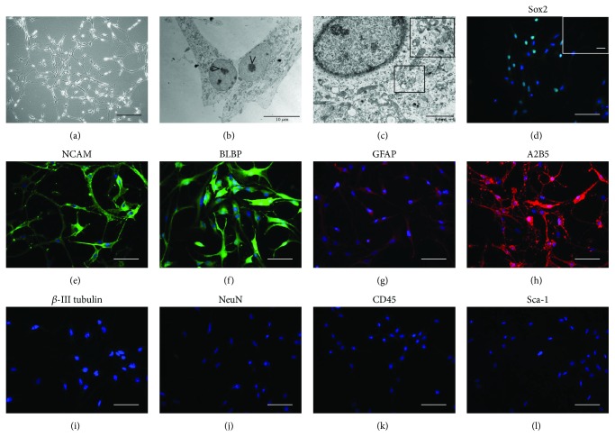
NSC characterization. NSC cultures were characterized by a large perikaryon and intercellular extensions (a). Ultrastructurally, NSCs were characterized by a large nucleolus (b) and by a cytoplasm rich in MVBs (c, insert). NSCs showed immunoreactivity for the NSC markers Sox2 (d), NCAM (e), BLBP (f), GFAP (g), and A2B5 (h). No reactivity was observed for β-III tubulin (i), NeuN (j), CD45 (k), and Sca-1 (l). Images show representative micrographs of NSCs isolated from 6 different foetuses. Scale bars: a: 200 μm; b: 10 μm; c: 2 μm. Scale bars: d–l: 50 μm.

