Figure 5.
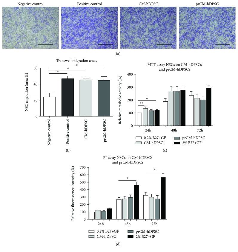
The effect of CM-hDPSCs and prCM-hDPSCs on NSC chemoattraction, metabolic activity, and proliferation. Transwell inserts stained with 0.1% crystal violet to demonstrate CM-hDPSCs and prCM-hDPSCs induced NSC chemoattraction (a) and showed that both CM-hDPSCs and prCM-hDPSCs (n = 4) attracted NSCs, but priming did not significantly increase this effect (b). The effect of CM-hDPSCs and prCM-hDPSCs (n = 6) on NSC metabolism and proliferation was evaluated by means of an MTT and PI assay, respectively. CM-hDPSCs and prCM-hDPSCs did not significantly influence NSC metabolism (c) or proliferation (d) after 24 h, 48 h, and 72 h although CM-hDPSCs significantly stimulate NSC metabolism after 24 h. No significant difference could be observed between CM-hDPSCs and prCM-hDPSCs. ∗p value ≤ 0.05 and ∗∗p value ≤ 0.01. Data are expressed as mean ± SEM. Scale bars: a: 200 μm.

