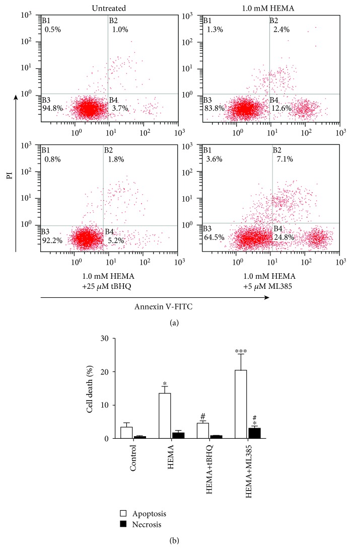
Figure 3.
Induction of apoptosis and necrosis in HEMA-exposed hDPCs. After a 24 h exposure period, cells were stained with Annexin V-FITC (Annexin)/propidium iodide (PI) and analyzed by flow cytometry. (a) Percentages of viable cells (unstained, B3), and cells in apoptosis (Annexin, B4), late apoptosis (Annexin & PI, B2) and necrosis (PI, B1) of one typical experiment are denoted in the quadrants of each density blot. (b) Bar graphs represent the mean values of flow cytometry data. Data represent mean ± standard deviations (n = 3). ∗P < 0.05, ∗∗P < 0.01, and ∗∗∗P < 0.001 vs. untreated cells (control group); #P < 0.05, ##P < 0.01, and ###P < 0.001 vs. HEMA-treated cells. Data were analyzed using one-way analysis of variance (ANOVA) and post hoc Tukey test.

