Figure 3.

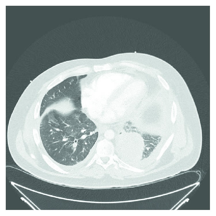
Computed tomography of the chest from June 2017 showing a left lower lobe opacity with preseptal thickening and a small pleural effusion.

Computed tomography of the chest from June 2017 showing a left lower lobe opacity with preseptal thickening and a small pleural effusion.