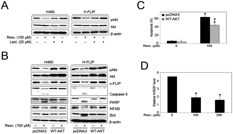
Fig. (6).
Akt overexpression inhibits resveratrol-induced apoptosis. (A) H460 and FLIP cells were pre-treated with lactacystin (proteasome inhibitor) for 1 hour before treatment with resveratrol (100 μM) for 12 hours and analyzed for pAkt and Akt protein expression by Western blotting. All blots were reprobed with β-actin antibody for loading control. Representative blots from three or more independent experiments are shown. (B) H460 and FLIP cells were transfected with pcDNA3 control and WT-AKT plasmid followed by resveratrol (100 μM) treatment for 12 hours, and analyzed for pAkt, Akt, c-FLIP, caspase-8, PARP, NFkB, and Bid protein expression by Western blotting. All blots were reprobed with β-actin antibody for loading control. Representative blots from three or more independent experiments are shown. (C) FLIP cells were transfected with pcDNA3 control and WT-AKT plasmid followed by resveratrol (100 μM) treatment for 12 hours and analyzed for apoptosis by Hoechst 33342 assay. Graphs represent relative fluorescence intensity over untreated control. Data are mean ± S.E.M (n≤4). *, p < 0.05 versus non-treated control. #, p < 0.05 versus resveratrol (100 μM) treatment. (D) FLIP cells were treated with resveratrol (0–200 μM) for 48 hours and VEGF levels were assessed by ELISA. Graphs represent relative fluorescence intensity over untreated control. Data are mean ± S.E.M (n≤4). *, p < 0.05 versus non-treated control.

