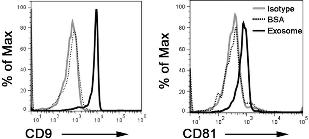
Figure 5. Exosome characterization by flow cytometry.

Exosomes (10 ug) were isolated by C-DGUC and non-specifically captured on latex beads, immune-stained and analyzed by flow cytometry. Flow cytometry dot plots show positivity for exosomes markers CD9 and CD81.
