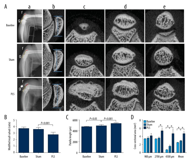
Figure 2.
Imaging findings of the patella baja created by patellar ligament shortening (PLS) surgery in the rat model of patellofemoral joint osteoarthritis (PFJOA). (A) X-radiograph images of the patella baja and changes of patella structure induced by PLS surgery. a) Radiographies of the lateral right knee in approximately 90° flexion with a specific right-angle device. b) X-radiographs of the sagittal length of the patella. c), d), e), representative cross-sectional micro-computed tomography (micro-CT) images at 4500 μm, 2700 μm, and 900 μm distal from the proximal region of the patella, respectively. f) The length of the patellar articular surface. g) The distance from the inferior edge of the patella articular surface to the end of the patella tendon. (B) The modified Insall-Salvati (MIS) ratio. (C) The patella length. (D) The analysis of cross-sectional area at different locations distant from the proximal region of the patella. Data are expressed as the mean ± standard deviation (SD). * P<0.05 versus the baseline group. # P<0.05 versus the sham group.

