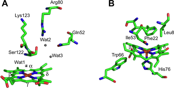
Fig. 3.

Overall fold of the canonical HemO of P. aeruginosa and IsdI of S. aureus. (A) HemO shown with heme in red and active site hydrogen bond contributing residues shown in stick form. (B) IsdG shown with heme in red and active site residues in stick form. Protein Data Bank (PDB) codes 1SK7 (HemO) and 3LGM (IsdI).
