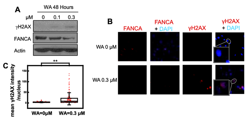
Figure 3. WA treatment results in accumulation of DSBs.

(A) DSB marker γH2AX increases along with WA treatments. FANCA is a replica of the corresponding gel in Fig. 2A. (B and C) Confocal microscopy analysis of immunofluorescence staining reveals elevated γH2AX in 0.3 μM WA treated nucleus (B) with statistical significance (C, ** p<0.01). Intensity of γH2AX fluorescence is quantified by using imageJ with DAPI masks.
