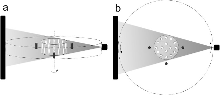
Figure 1.
Geometric setting of the radiographic phantom in the CBCT unit: (a) lateral view; (b) axial view. The area between the full and dotted lines represents the exomass. The black rectangle and the shaded triangle represent, respectively, the image receptor and the X-ray beam. CBCT, cone beam CT.

